Abstract
The aim of this study was synthesis and characterization of different kinds of nanoparticles such as gold nanoparticles (GNPS) and rare earth elements-doped upconversion nanoparticles (UCNPs) and amplification of wild type M13 phage. M13 wild type phage was amplified and characterized by Atomic Force Microscopy. Characterization of these nanoparticles is done via Ultraviolet-visible spectroscopy which depicts absorption in the ultraviolet-visible spectrum, TEM and AFM. Gold nanoparticles in the size range of 10- 20 nm were synthesized with and characterized by Transmission Electron Microscopy (TEM) showing excellent spherical morphology well within the size rage. Upconversion nanoparticles were synthesized and observed through TEM depicting rod like particles. Bright green fluorescence was observed with a NIR laser showing UCNPs were successfully synthesized. One of the most widespread cancers in the human body is breast cancer which also has the highest survival rate. The goal is to synthesize nanoparticles bound with phages that can be targeted towards breast cancer therapy and diagnostics.
Introduction
Nanoparticles have been the focus of study due to their size and shape dependent physicochemical properties which have several applications in catalysis, photonics and biomedicine. They have a large surface area as well as high fluorescent intensity which allow them to be used for detection methods in diagnostics and therapy. Their surface properties are complicated but can be altered for purposes such as drug delivery and ligand binding. Gold nanoparticles have been under study as model drug delivery platforms because of their surface properties and low toxicity which allows convenient functionalization with chemical and biological molecules. Surface Plasmon resonance makes GNP suitable for bioimaging, biomedical therapeutics, and biodiagnostic tools.1 Upconversion nanoparticles have the ability to convert near IR radiations with low energy into visible radiations with high energy. They have progressed as alternative fluorescent labels giving them an edge to traditional fluorophores. They are employed for imaging and biodetection assays in both in vivo and in vitro applications.2
Phages are viruses that specifically infect bacteria. They are sorted according to the structure of there capsids withholding the genomes, chemical and structural composition of their genomes, and the mechanism of mRNA production.3
One particular challenge faced with nanoparticles is their assembly into well constructed structures. Nature provides biomolecules such as nucleic acids, proteins, and viruses which can be used to integrate the nanoparticles. The advantage of combining the nanoparticles with phages and form a nanoparticles-phage hybrid has led to nanostructure organizations with controlled shape, size, alignment and orientation.4
Background
Researchers have come up with various methods of developing gold nanoparticles which can be used for different treatment purposes. They are essential since they can vary in size, shape and structure. The seeded growth strategy for the synthesis of large citrate gold nanoparticles based on controlling the size and shape is described by the inhibition of secondary nucleation throughout the homogenous growth process which allows the magnification of Au Nps via the reduction of AU3+ by sodium citrate. The growth process is monitored by controlling the temperature, pH level, and seed concentration. This allows for the particles with higher monodispersity and for enlargement of smaller predetermined size particles.1 Different kinds of gold nanoparticles shapes can be synthesized. Gold nanospheres (AuNPs) are synthesized by the reduction of chloroauric acid resulting in solid spheres made of gold ranging in diameters from a few to 100 nm. Gold nanoshells (AuNSs) are also spherical in structure with an internal silica core and thin layer of gold. They can range in size from 50-150 nm.5 Gold nanorods (AuNRs) are developed from choloroauric acid with a gold seed and a stabilizing agent (CTAB).6 AuNRs range typically in the size of 25–45 nm in diameter. Gold nanoparticles such as AuNRs or AuNSs have optical properties of light absorbance and scattering in near-infrared (NIR) wavelengths (650–900 nm). Two other forms of gold nanoparticles are hollow gold nanospheres. The modifications in size, shape and surface properties of gold nanoparticles can therefore be used in the treatment of many types of cancer, in specific, breast cancer. Studies have shown that gold nanoparticles ranging from 4-10 nm in diameter were easily absorbed and found and accumulated in organs such as the liver, kidney, spleen and brain.7 The clearance of these nanoparticles from the body depends upon the surface charge and size. The ability to synthesize spheres, shells, rods and cages of varying sizes and shapes makes gold nanoparticles more advantageous as compared to other non-metallic nanoparticles such as liposomes.
Lanthanide-doped upconversion nanoparticles (UCNPs) convert near infrared radiation (NIR) to visible light by converting two or more low energy excitation photons into shorter wavelength emissions. They are excited by NIR which makes them emit strong visible fluorescence. This makes them ideal with advantages due to their unique optical properties. Organic dyes and quantum dots (QDs) have multiphoton absosptions which makes UCNPs different from them. UCNPs are prone to depict sharp emission peaks, high photochemical stability, high resistance to photo bleaching and long emission lifetime. UCNPs take part in many applications such as bioimaging, biosensors, drug delivery and photodynamic therapy. Hydrophilic and biocompatible UCNPs are employed in both therapy and diagnostics (theranostics).8 For bioimaging, UCNPs produce virtually zero autoflourescence background, large anti-Stokes shift, narrow emission bandwidths and high resistance to photobleaching which make them best fit for repetitive imaging. Another important property is the low levels of light scattering which allows for deep tissue penetration because of excitation range being in the NIR region.9 UCNPs have also been widely used in the development of latent fingerprints due to their physical and chemical properties including small particle size, large surface area, high photochemical stability and high fluorescent intensity. Research has shown them to surpass conventional powders and other nanoparticles. Commercially available and used UCNPs include NaYF4:Yb,Er. One way to go about the synthesis NaYF4:Yb,Er UCNPs with uniform size and strong fluorescent intensity is to proceed with a solvothermal approach. The effects of reaction time, reaction temperature and volume of oleic acid on the size, phase, and UC fluorescence intensity of UCNPs need to be optimized. (REFERNCE: FINGERPRINT) .Together gold nanoparticles (GNPs) and upconversion nanoparticles (UCNPs) are being investigated in the field of theranostics.
There are two categories of bacteriophages present namely lytic and temperate phages. Lytic phages are prone to breaking open and killing the host bacteria after infection for release. In contrast, temperate phages use the host bacteria as “factory” for phage amplification and secreted without killing of the host. Some exception like the lambda phage can follow either the lytic or lysogenic cycle. They are employed in the delivery of plasmid DNA into mammalian cells without any levels of cytotoxicity. Professionally lytic phages are employed in antibacterial applications example the T7 phage. Filamentous phages like the Ff, M13, f1, fd, Ike and N1 are found to have more applications as compared to lytic phages as they are known to self-assemble. Phages can exist in diverse forms of shapes and sizes from long rod like filaments to more compressed shapes with much shorter tails. The versatility of their different shapes and sizes provide a powerful building material allowing them to be assembled as nanomaterials. Over the past years, nanotechnology has incorporated naturally occurring phages like the filamentous M13 for drug delivery, cancer treatment, imaging and tissue regeneration. Due to the natural processes of phage and the diversity in there structures, they are essential tools in nanomedicine for precision targeting and self assembled biomaterials. They are utilized for enhanced bioimaging for disease detection, nanotherapeutics for disease treatment, drug delivery, vaccination, stem cell differentiation (mainly bone regeneration) and tissue formation.3
Results and Discussions
GNP synthesis
Before addition of the gold reducing agent the gold is in the AU3+ form. When the sodium citrate is added, gold atoms tend to form in solution and end up with a red wine color (Figure 1). Nucleation is the process by which particles are formed. The remaining gold atoms bind to nucleation sites and growth occurs. The concentrated reducing agent tends to give smaller nanoparticles. Obtaining GNPs with a large size and stability depends on temperature of the solution, control of pH of solution and number of gold atoms injected in each step.1
In order to obtain GNPs with a controlled size, morphology and concentration, we adjusted the temperature of the solution. Particles synthesized at 90º C showed excellent morphology and narrow size distribution. UV-vis analysis was kept well within range of 400 nm to 600nm.UV-vis analysis showed highest absorbance peak at wavelength of 529 nm (graph 1). To determine the size and morphology the GNPs were analyzed with the TEM. The nanoparticles were spherical in shape with slight aggregation. The size range was between 10nm and 20 nm and the mean diameter of the nanoparticles was 10 nm (figure 2).
In another study, Au Nps were grown from a size range of 30 nm to 200 nm. Au NP size measured from TEM images that growth of AuNPs was not homogenous and the size decreased. This shows that growth is determined by relation between diameter of growth particles to amount of gold added.1

Figure 1 Synthesis of Gold Nanoparticles showing red wine color.

Graph 1: UV-vis analaysis of Gold nanoparticles showing highest wavelength peak at 529 nm.
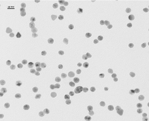
Figure 2 TEM image of Gold Nanoparticles.
UCNP synthesis
Rare earth NaGdF4:Yb,Er upconversion nanoparticles were synthesized using a solvothermal approach. The size and morphology of the NaGdF4:Yb,Er NPs were characterized by TEM (figure 3). The TEM image shows UCNPS of similar morphology. They are in the mixed phase consisting of of spherical shaped particles. Slight aggregation of particles is depicted by the dark colors. Some of the UCNPs showed stacking on top of the other showing uniformity with a size of 50 nm approximately. The NaGdF4:Yb,Er NPs exhibit bright green upconversion luminescence under 980 nm as seen in figure 4. Three visible emission spectra showed peaks at wavelength between 100-200 nm, the second at 270 nm and the highest peak at 580 nm different sizes (graph 2). Research has shown that nanoparticles excited by the NIR laser at 980 nm for cellular and molecular imaging allow deep penetration into tissues and is also less harmful.10
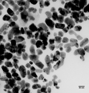
Figure 3 TEM image of UCNPs

Graph 2: Fluorescence spectrometry showing 3 peaks at different wavelengths observed for UCNPs.
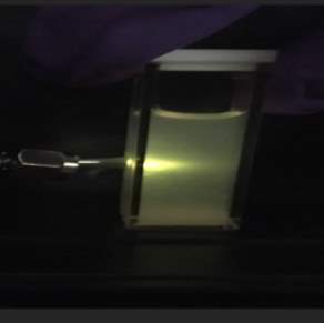
Figure 4 NIR laser shown through UCNPS depicting bright green fluorescence.
Phage amplification

Figure 5 TEM image of M13 Wild type Phage
Wild type M13 phage possesses a single stranded DNA (ssDNA) genome packed into a protein coat arranged with 2700 copied of the major coat proteins (pVIII) and five copies of each minor coat protein (including pIII, pVI, pVII, and pIX).(REEFERENCE). The ssDNA encodes 11 proteins which include five structural coat proteins. The amino terminus of each pVIII protein is exposed to the surface away from the phage. Phages were harvested from 1L of LB culture media. To determine the morphology of the phages, the phages were observed under AFM. The M13 phage has a flexible rod like shape. The phage solution was also analyzed using UV-Vis Nanophotometer. The absorbance peak was at a wavelength of 269 nm which was used to calculate the concentration of about 0.042 x 10⁻¹³.
Studies have comparatively analyzed methods better suited for determining phage concentrations efficiently and reliably. Traditional Plaque Assay (PA) has always been the standard for quantification of phages but produces results over a longer period of time whereas using a Nanophotometer requires expensive equipment but the results generated are much faster and less exhausting.11
Summary and conclusions
Main conclusions of the study should be presented. Conclusions must be based on a critical analysis of the results and discussions presented earlier. If appropriate, state how the conclusions point to future investigations.
The synthesis of citrate-stabilized gold nanoparticles well within the size range of 10-20 nm brings about their well-deserved relevance to biological and biomedical applications. Their conjugation with biomolecules such as peptides, antibodies and DNA makes them efficient drug delivery carriers. They can also be used as model nanoparticles systems to study the effect of nanoparticle morphology on biological systems.
UCNPs with distinct hexagonal shapes synthesized can advance in the world of bioimaging or drug delivery as nanocarriers and photodynamic therapy. The solvothermal approach of synthesizingNaGd4:Er,Yb led to improved fluorescence intensity under NIR irradiation. They can be used as fluorescent labels with higher sensitivity, contrast and low background interference as compared to traditional fingerprints.
The ability of phages to display molecules such as nanoparticles and other peptides on their surface allowed them to use in fields such as disease diagnosis and treatment, tissue regeneration, bacterial and fungal infection detection, vaccination, and gene therapy.
Materials and Methods
Materials:
Phage amplification
LB medium, Sodium chloride (NaCl), Polyethylene glycol (PEG) was purchased from VWR. Tetracycline was purchased from Sigma Aldrich. E.Coli ER2738 was purchased from NEB. Wild type phage M13 was also purchased from NEB.
Synthesis of gold nanoparticles:
HAuCl4.3H2O (99%) and trisodium citrate (99%) were purchased from Sigma-Aldrich. Milli-Q water was used in all experiments. All glassware was cleaned with acetone, rinsed with deionized water and stored at 150֯ before use.
Synthesis of upconversion nanoparticles:
Rare earth oxides used in this work included gadolinium nitrate (Gd(NO3).6H2O), ytterbium oxide (Yb2O3), erbium oxide (Er2O3) were all 99.99% purity and were purchased from Sigma-Aldrich. Sodium fluoride (NaF), Oleic acid (C13H33COOH), stearic acid (C17H35COOH) were also purchased from Sigma Aldrich. Sodium hydroxide (NaOH), Nitric acid (HNO3) were purchased from FISCHER.
Methods:
M13 wild type phage amplification
1 L LB medium culture of M13 wild type phage was carried out over a period of 5 days.
Day 1: 1L of LB medium was prepared and autoclaved in the morning. 5 ml of LB medium was inoculated with the E.Coli ER2738 after and 5 µl of tetracycline. The culture was incubated under 37 ºC at a speed of 200-220 rpm overnight.
Day 2: in the morning we transferred 200 µl of the overnight culture and 20 µl of stored wild type M13 phage solution to 5 mL of LB medium (in a test tube) and incubated for 4-6 hours under 37 ºC at a speed of 200-220 rpm. In the afternoon we transferred 500 µl of the culture into 1 L of LB medium and incubated for 18 hours under 28 ºC at a speed of 200-250 rpm
Day 3: In the morning, we centrifuged the culture under 4 ºC at 8200g for 40 min. we then transferred the supernatant into a bottle, added 40g PEG 8000 and 30g NaCl, and incubated at 4 ºC overnight.
Day 4: the supernatant PEG/NaCl was centrifuged under 4 ºC at 8200g for 50 min, the white pellet was retained (phage pellet), and centrifuged under 4 ºC at 8200g for 5 min again, the rest of the supernatant was removed using pipette.
We added 60 mL of water to the bottle to dissolve the phage white pellet by pipetting the solution up and down, transferred the 60 mL of phage solution into 2 centrifuge tubes and incubated at room temperature with gentle shaking for 1 hour.
We centrifuged the phage solution under 4 ºC at 11500 rpm (rotor 13.1) for 30 min, transferred the supernatant to two new centrifuge tubes, add 0.3g PEG 8000 and 0.22g NaCl to each tube and left to incubate for 1 hour on ice.
We centrifuged the phage solution under 4 ºC at 7500 rpm (rotor 16.250) for 30 min, transferred the supernatant to two new centrifuge tubes, add 0.3g PEG 8000 and 0.22g NaCl to each tube and left to incubate overnight.
Day 5
We centrifuged the phage solution under 4 ºC at 7500 rpm (rotor 16.250) for 30 min, dumped the supernatant, centrifuged under 4 ºC at 7500 rpm for 5 min again, removed the rest supernatant solution using pipette.
The phage white pellet was dissolved with 10 mL of water
Synthesis of gold nanoparticles
Gold nanoparticles were synthesized using a standard protocol. 120 ul of 60mM HAuCl4 was added to a beaker with a magnetic stir bar on a hot plate and left to bring to a boil for 10 minutes. The RPM was set to 600. The temperature was set at 95º C. To the rapidly-stirring boiling solution, 1.5 ml of 640 mM sodium citrate was added. The solution color changed from pink to red wine color which is the color of the gold nanoparticles.
Synthesis of NaGd4:Er,Yb upconversion nanoparticles12
1. Synthesis of Rare-Earth Stearates Precursors: Rare-earth stearates were used as a precursor. To synthesize the rare-earth stearates, a mixture of 0.3941 g of Er2O3, and 0.0383 g of Yb2O3 was first dissolved in nitric acid followed by heating. 0.8807 g of Gd(NO3).6H2O was also mixed. The resulting nitrate powder was obtained after the solvent was removed by drying. The as-prepared powder and 8.5344 g of stearate acid were dissolved with stirring in 80 mL of ethanol at 78 °C in a flask. Another solution containing 1.1900 g of NaOH and 20 mL of ethanol was added drop wise to the flask within 30 min. The resulting mixture was then refluxed at 78 °C for 40 min. Precipitates from the reaction mixture were filtered under decompression and washed, first with water twice and then with ethanol once. The precursor powder was obtained after the precipitates were dried at 60 °C for 12 h.
2. Synthesis of NaGd4:Er,Yb UCNPs by a Solvothermal Approach— Mixing of 10 mL of water, 15 mL of ethanol, and 5 mL of oleic acid under stirring resulted in a homogeneous solution, to which the precursor powder (0.9578 g) and NaF (0.2099 g) were added. The resulting mixture was sonicated for 15 min, transferred to a 50 mL autoclave, sealed, and then solvothermally treated at 180 °C for 24 h. After air-cooling of the autoclave to room temperature, UCNPs were deposited at the bottom of the vessel and then collected by a mixture of chloroform and ethanol (1:6, v/v). The UCNPs were purified by centrifugation, washed twice with a mixture of water and ethanol (1:2, v/v), and then dried at 60 °C for 12 h. UCNPs were thus obtained.
Characterization:
UV/Vis Analysis:1, 3
Gold nanoparticles and phage solutions were analyzed using an IMPLEM NanoPhohtometer. The instrument scans between 200 and 950 nm. The control used was air with no cells present during the background scan. Thus a UV/Vis spectrum was obtained. AuNP solution: Approximately 1 ml of solution was pipetted into a 1.5 ml plastic cuvette with additional 1 ml MilliQ water. The scan performed was in the range 400 and 800 nm. The mode was set to absorbance.
Phage solution: Approximately 1 uL was pipette into LabelGuard Microliter Cell Nanodrop with a factor 10 lid. The scan was performed between 200 and 950 nm, but the absorbance at 296 nm was used to calculate the concentration of the phage solution using Beer’s Law.
TEM:9
TEM was performed on UCNPs using a ZEISS10A with an accelerating voltage of 80 kV. A ten L droplet of each sample was placed on the carbon side of a 400 mesh copper TEM grid and left to air dry (about 30 minutes). Samples were imaged and used for size measurements and visualizing aggregation and distribution.
Fluorescence Spectrometry:
Fluorescence spectra were acquired for UCNPs using an Agilent Cary Eclipse
Fluorescence Spectrometer closed off to room light. UCNP solution (~1 mL) was loaded into a 3 ml quartz cuvette cell and its fluorescence spectra analyzed using chemiluminescence mode regarding the emission wavelength. Due to the limitation of this instrument allowing sensitivity up to 900 nm (red shift), external continuous-laser integration was used to allow for an exact excitation wavelength of 980 nm. The external 30W 980 nm VD VII DPSS LASER DRIVE was set at 1.3 W.
AFM:12
Phage images were produced using a Bruker BioScope Resolve AFM. ScanAsyst Air probes were used on ScanAsyst mode during scanning. The laser was aligned in a way that probe could engage with the sample surface. Feedback was optimized and the sample was scanned. The probe was adjusted to obtain the image in a desired area of the sample.
Acknowledgments
I am very grateful to Dr. Mao for allowing me to learn and gain experience and knowledge in his lab. I would like to thank Gabbrielle Abbot for teaching me all the skills at hand and putting her time and effort into helping me gain sufficient knowledge in this field. I would also like to thank MengMeng for investing her time for the experimental procedure.
Supplementary materials
Figure 6 GNP DLS

Figure 7 GNP DLS
|
Collection Time: 12/12/2016 11:51:25 AM |
|
|
Operator Name : |
|
|
Scan Software Version: 1.2(147) |
|
|
Parameter List : |
|
|
Instrument Cary Eclipse |
|
|
Instrument Serial Number MY13160003 |
|
|
Data mode Bio/Chemi-luminescence |
|
|
Gate time (ms) 5.0000 |
|
|
Scan mode Emission |
|
|
X Mode Wavelength (nm) |
|
|
Start (nm) 400.00 |
|
|
Stop (nm) 1000.00 |
|
|
Em. Slit (nm) 5 |
|
|
Data interval (nm) 1.0000 |
|
|
Emission filter Open |
|
|
PMT voltage (V) 1000 |
|
|
Corrected spectra OFF |
|